In 2019, popular Bollywood actress Disha Patani was practising gymnastics on her terrace when she fell on the concrete floor, injuring her head. The head injury caused short-term memory loss as she could not recall anything from six months before the incident.
Head injuries are tricky as they can damage the spongy inner brain. Depending on where the brain gets hit, functions like speaking, chewing, breathing, memory, and walking get affected. An earlier article covered how a head injury can cause traumatic brain injuries and the complications that arise due to them.
Catch it early
Although we can do our best to avoid head injuries, what happens when luck is not in our favour?
At such times, immediate medical attention is vital, and assessing the degree of injury to the brain is crucial for better treatment and quick recovery. Dr N K Venkataramana, the founder of BrainS Super Specialty Hospital, Bengaluru, says that treatment for head injuries starts with checking the affected person’s ABCs — airway, breathing, and circulation. “Individuals who meet with an accident should have a clear airway, proper breathing, and blood circulation,” he says. He further adds that the doctors check for consciousness and disabilities followed by scanning for unseen damages like spinal and brain injury.
Dr Venkataramana and Ashutosh Vaish, a researcher at Indraprastha Institute of Information Technology, Delhi, give insights to Happiest Health about the scanning methods currently being used to assess damage in a head injury.
-
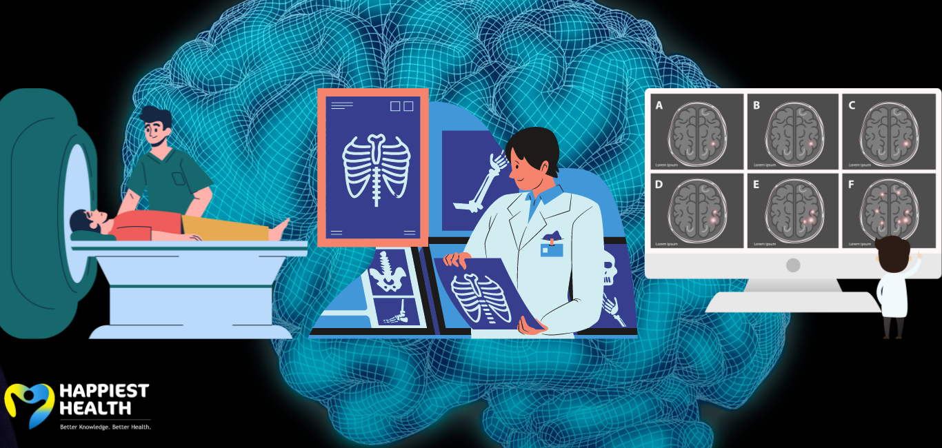
The X-ray factor
Wilhelm Roentgen’s revolutionary discovery of the X-ray set the base for all scanning methods. An X-ray gives a physician a visual of the damage, such as a broken bone or a thin crack in the skull. However, taking an X-ray requires the injured to be conscious to explain where the pain is felt, like in the neck or the back. “It is essential to do an X-ray for a spinal injury because it is not seen outside,” says Dr Venkataramana.
In this technique, X-ray beams travel through the body and are absorbed in different amounts by different tissues, depending on the nature and type of tissues they pass through. The black and white film can detect fractures, infections, and bone defects in the skull.
-
Seeing the impaCT
“Computed tomography or CT is an extension of the X-ray,” says Vaish, adding that it allows the physician to assess the details of the internal damage. This technique collates multiple X-ray images in 2D format taken from different angles around the body and is processed on a computer. The computerised scan is sharp, clear, and three-dimensional, which helps to distinguish between different types of tissues and injuries, thus giving an upper hand over the standard X-ray system.
Dr Venkataramana says the CT scan is the most preferred technique for screening brain injuries. “A CT scan of the brain can show the various types of injuries starting from compound fracture, blood clots, contusions, haemorrhages, and brain swelling,” he explains.
-
The magnetic hummer
Along with CT, magnetic resonance imaging (MRI) is also the gold standard imaging technique for assessing brain injuries.
Dr Venkataramana prefers the CT over the MRI. However, Vaish is in favour of the MRI as it gives a detailed picture of the brain’s structural organisation.
An MRI scan is recommended when the symptoms following a brain injury persist for more than 48 hours, Vaish says. It is taken by passing the person through a large tube-shaped magnetic chamber. The imager comprises both magnetic and radio waves. The strong magnetic field aligns the water molecules in the body through which the radio waves pass to get a clear and detailed image of organs and tissues.
“An MRI helps you to know whether the blood is flowing correctly, or which part of the brain is affected due to clot or toxic proteins,” says Vaish. This technique is also used in detecting stroke and neurodegenerative diseases, he adds.
-
Tech 2.0- diffusion MRI
Vaish explains that conventional scanning methods show the overall structural damage but not minute tissue damages in the brain. When minute tissue-level details are required, a modified version of the MRI—diffusion MRI — is useful. “It is an advanced and emerging technique that measures the movement of water molecules within the tissues. The MRI machine detects this movement and represents the changes as different levels of brightness in the resulting image,” says Vaish.
Other advanced neuroimaging techniques like positron emission tomography, electroencephalography, magnetoencephalography, functional MRI and functional near-infrared spectroscopy give intricate architectural details of the brain. However, they are used more in laboratories for molecular investigations of the brain.
Editor’s note: 20 March is World Head Injury Awareness Day. This article is the second in a two-part commemorative series.