Ureteropelvic junction (UPJ) obstruction is a condition where there is a partial or complete blockage in the area that connects the renal pelvis (part of the kidney) to the ureters (tubes) that carry urine to the bladder. While this is not a common condition in the children and occurs only in 1 in 100 babies, congenital ureteropelvic junction obstruction can lead to serious conditions like hydronephrosis (a condition where one or both kidneys become swollen due to the buildup of urine). These conditions can cause kidney damage in children if timely diagnosis and treatment is not given.
Prenatal case of ureteropelvic junction obstruction
Two weeks ago, I came across an eight-week-old female baby from Bangalore who had been diagnosed with ureteropelvic junction obstruction (a condition where a block had formed between the baby’s kidneys and ureter). Due to this, the urine flow was partially affected. The child’s parents were worried. On enquiring with the parents, I learnt that the condition was initially diagnosed during the routine ultrasound examination performed on the mother as part of the antenatal care. But the parents did not know how to deal with the situation.
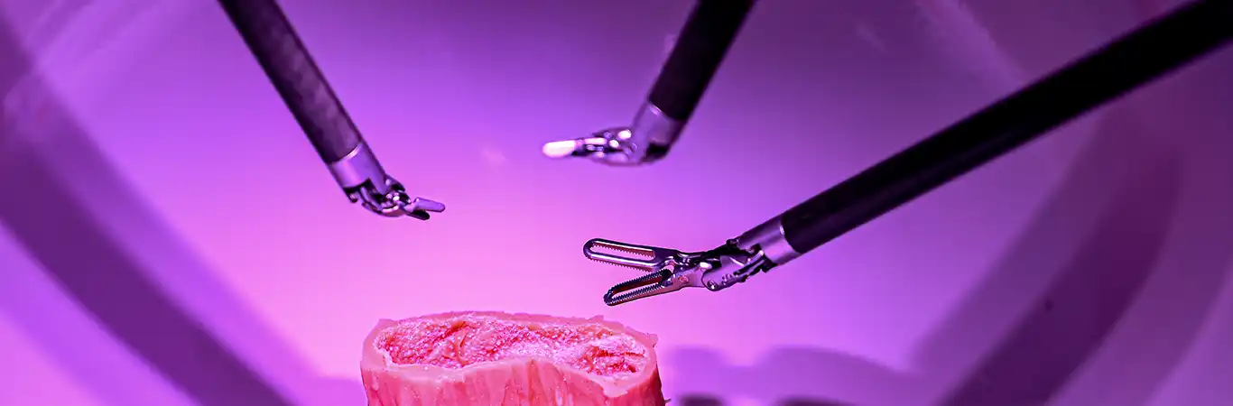
As the ureteropelvic junction obstruction was significant, we decided to conduct pyeloplasty surgery (removal of block from the kidneys). While there is no medical therapy available to treat the UPJ obstruction, this condition is now being increasingly diagnosed in new mothers during ultrasound examination. This can help us in giving timely treatment to the children.
The surgery was successful, and the block in the baby’s kidneys was removed without further damage. The kidneys have started functioning normally and the baby is currently recovering from the surgery. A follow-up will be done soon to check the progress in the baby’s condition.
Postnatal case of ureteropelvic junction obstruction
In another case, a six-year-old male child from West Bengal along with his parents consulted me with severe pain in his abdomen. Without further delay, a renal ultrasound was performed to detect the actual cause of the child’s pain. The ultrasound revealed that the child was having ureteropelvic junction obstruction causing severe hydronephrosis.
This six-year-old boy was suffering from this condition for a long time. His abdomen had been partially occupied due to the enlargement of kidneys, causing severe pain and discomfort. Further diagnosis revealed that his kidneys were functioning only at 19 percent, affecting his overall kidney health. Though these abnormalities are easily detected during the foetal ultrasounds, the parents were unaware about the condition during their pregnancy. There were no symptoms of hydronephrosis after his birth, too, they shared.
Since the functioning of the child’s kidney was affected, a pyeloplasty surgery was performed. A ureteral stent was also placed to treat hydronephrosis and the child was discharged in a healthy condition.
While he is recovering, a follow-up procedure will be done later to remove the stent, which was placed during the surgery. We will also check for any dilation of the kidneys through an ultrasound scan. Therefore, early treatment is important.
As told to Steni Simon
The author is the head of the department-pediatric surgery and pediatric urology at Manipal Hospitals, Bangalore.