Growing a functioning heart in a laboratory may seem like a far-fetched idea, but scientists have made significant strides in achieving just that. Heart organoids or “mini hearts” are not as complex as the real thing. However, they mimic the beat of a human heart and offer a unique opportunity for researchers to study the heart’s development and response to medications and treatments.
In a recent study, a team at the Technical University of Munich (TUM) successfully created heart organoids that emulate the earliest phases of heart development. These heart organoids grow as small balls of cells in a lab dish, allowing researchers to observe their development and function in a controlled environment.
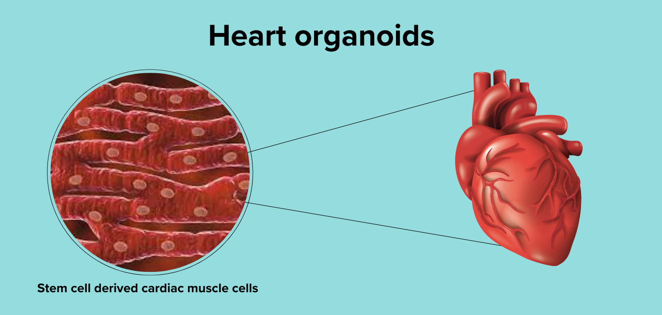
Stem cells unlock heart research
In the past, scientists have used animals like mice to study the heart and its conditions. However, animal models have proven to be highly inaccurate when it comes to modelling the human heart.
“Results from animals cannot always be translated to humans. Being able to study the function of human cardiac cells in the lab will accelerate the investigation of disorders such as congenital heart malformations,” lead author of the study Alessandra Moretti, a professor of regenerative medicine in cardiovascular disease at TUM, said.
One of the key technical advances that made it possible to grow these organoids is stem cells which can change into any cell in the body. So far, while stem cells were used in the study of many other organs in the body, the heart has always posed a challenge owing to the different cell types that make it up and its complex architecture.
Moretti owes this complexity to the different pathways that cells take during the heart development process, one that involves the coordination of multiple cell types at a time during embryo development.
Her team was the first to obtain heart organoids with the epicardium (muscles that line the inner walls of the heart). “The epicardium plays several important roles during development, which is why it is useful to understand its ontogeny (developmental history),” Moretti said in an email exchange with Happiest Health.
The team devised a protocol that used pluripotent stem cells to form the heart organoid which they are calling an ‘epicardioid’. The epicardioids consist of a ball of 35,000 cells that have formed over the period of a few weeks by adding different signalling molecules to it.
“In this way, we mimic the signalling pathways in the body that control the developmental program for the heart,” explains Alessandra Moretti.
The results of their work have been published in Nature Biotechnology.
Also read:
Stem cell based synthetic mini organs for parathyroid treatment
How and when 3D printed organs may become a reality
Uncovering the basis of heart conditions
While this work has been able to give researchers key insights into how the heart develops, it has also been a useful tool to study heart conditions.
Using pluripotent stem cells from an individual suffering from Noonan syndrome (a genetically inherited disorder that can cause defects in many organs in the body including the heart), they were able to emulate the characteristics of the condition in the lab.
“We believe that epicardioids will be particularly helpful for the study of congenital heart diseases caused by gene mutations. They could also become useful tools for testing new drugs or gene-based therapies,” says Moretti.
Additionally, Moretti is also interested in learning about heart regeneration using the epicardioids.
“The adult human heart has almost no capacity for regeneration, which is what makes heart attacks so dangerous,” she says.
She says that certain species of lower vertebrates such as the zebrafish, are capable of rebuilding adult heart muscle after injury – where they have observed the epicardium to be central to this process.