Just as the king on a chessboard is protected against potential threats from all sides, the brain also has a tight security system to prevent a check-mate situation. One of these protector pawns is the blood-brain barrier (BBB), which also mirrors functional changes inside the brain, although the exact mechanism by which it does so is unclear.
Understanding how the BBB responds to these changes requires an in-depth cellular picture; however, a lack of real-time imaging methods has so far limited scientists from visualising them.
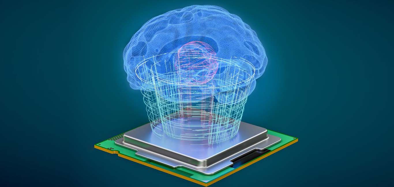
Not anymore. Scientists from the University of Bonn, Germany, have developed a novel technique that provides greater insight into the brain’s gatekeeper, which could help in decoding neurological disorders.
“Looking closely at the blood-brain barrier in different pathological settings has become increasingly essential not only to understand the pathogenesis of a disease but also to design therapeutics accordingly,” Dr Amira Hanafy, the study’s first author and a postdoctoral fellow at UKB’s Clinic of Neurosurgery (University Hospital Bonn), tells Happiest Health.
The brain’s gatekeeper
The BBB comprises a network of various types of cells interwoven with blood vessels forming a tight cellular mesh that restricts the entry of even water and small molecules. Studies have found that sleep, stress and nutrition can compromise the barrier’s integrity, causing leaks. The architectural changes probably alter neuronal activity – often the culprit behind neurological conditions.
“It has been evidenced that a handful of neurological diseases involve blood-brain barrier alterations such as Alzheimer’s disease, Parkinson’s disease and Tourette’s syndrome,” Dr Hanafy says.
Read more: Decoding the genetics of Alzheimer’s: the role of APOE4 and SHMOOSE
While studies could identify some of the pathways leading to changes in neuron activity, some others are not fully understood. Although several lab models of BBB have been developed in recent decades, each has its own merits and demerits, adds Dr Hanafy.
For example, how the BBB responds and adapts dynamically to different test substances added to the intravascular fluid is not entirely clear yet. The described model along with the imaging technique could help understand how medicines work a live-functioning scenario.
Understanding the barrier
The present study, published in Nature Communications, explains these details. Prof Dirk Dietrich, the study’s lead investigator, provides the example of a flat tyre to demonstrate their technique. “If the tyre loses air, you don’t know where the leak is. That is why you hold the inflated bicycle tube under water to identify the leak. This principle also underlies our method,” he says in a statement.
The team built their model from the brain tissues of mice and human participants with seizures.
The primary goal was to keep the sliced brain tissue alive (6-8 hours) to monitor the activities of the BBB. They tweaked a known electrophysiology technique called microperfusion to study how the fluids interact with the BBB cells.
The scientists kept the tissue in a solution that mimicked the cerebrospinal fluid or the internal conditions for the barrier, Dr Hanafy explains. They also filled the blood vessels with liquid via microperfusion to see how the cells responded.
“We pierced a blood vessel within the alive brain slice and introduced a solution with external pressure while monitoring the interaction of said solution with the blood-brain barrier within the blood vessel,” she adds.
The scientists scanned the fluid with an advanced instrument called a multiphoton microscope that could trace potential leaks in the blood vessels. The scanning method employs light particles that penetrate deep into the tissue, allowing the scientists to obtain a clearer picture of the cellular happenings.
“The technique can reveal blood-brain barrier disturbances and assess neuronal activity,” says Dr Hanafy.
Looking ahead
Dr Hanafy is optimistic about the future of understanding the blood-brain barrier, “We used our novel technique to identify some blood-brain barrier alterations in an Alzheimer’s disease mouse model,” she says. Her team is also investigating how the blood-brain barrier develops and how its cellular structure fluctuates.