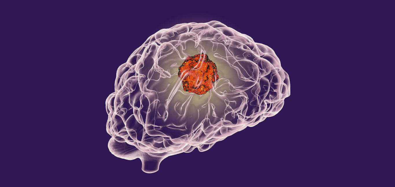
Brain tumours pose extreme challenges where treatment is concerned. The type of tumour, whether or not it is aggressive, and if yes, how aggressive it is are all gauged using biopsy (tissue samples). However, identifying types of brain tumour from biopsy results is time-consuming, time that is quite a significant factor in the lives of people who have the tumours. Scientists are actively exploring using AI (Artificial Intelligence) and ML (Machine Learning) models to address this concern and better understand brain tumours.
In one such attempt, researchers from the Harvard Medical School, USA, have developed an AI tool that can decode the tumour DNA to help understand the molecular makeup of brain tumours. This tool is awaiting FDA approval and could potentially help neurosurgeons take critical decisions during surgery.
A study published in the journal Med explains this breakthrough involvement of AI in treating brain tumours. Dr Kun-Hsing Yu, lead on the study and assistant professor of biomedical informatics at the Blavatnik Institute at Harvard Medical School, tells Happiest Health, “We spoke with several neurosurgeons, pathologists, and oncologists around two years ago, recognised the diagnostic challenges they faced every day and determined to design novel AI methods to solve their critical clinical problems.”
The AI tool that Dr Yu and his team ultimately came up with can give surgeons a real-time picture of the tumour tissue during brain cancer surgery. This can help them understand the aggression level of the tumour and decide how much of the cancerous brain tissue needs to be removed.
Need for evaluation
Surgery, radiation therapy, targeted therapy, and immunotherapy are the most practised treatment approaches to brain tumours. However, these methods still have drawbacks that prevent experts from adequately examining and giving accurate clinical evaluations of the brain tissue.
“The limitations of the current modality, which is called frozen section [tissue sample for biopsy], is that it cannot distinguish between the grade of tumours. We do not get 100 per cent accurate tissue diagnosis,” explains Dr Priyank Vasavada, a neurosurgeon at M S Ramaiah Medical College and Hospital, Bengaluru.
Read more: A step in understanding brain tumours
Read more: IIT Madras creates AI tool to identify glioblastoma
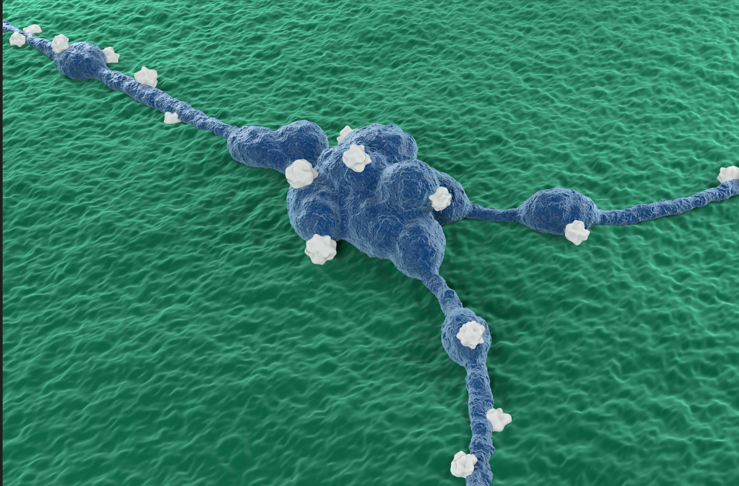
Cracking cancer’s molecular code
Using pathologists’ input, the researchers designed an AI tool called CHARM (Cryosection Histopathology Assessment and Review Machine). CHARM analyses the molecular structure of the various types of brain cancer.
To develop this AI tool, the researchers analysed 2,334 brain tumour samples from 1,524 people from different age groups who had glioma (cancerous brain tumour). To check the accuracy of CHARM, they tested the AI tool using a new set of brain tumour tissues. This tool achieved an accuracy of 93 per cent in distinguishing the tumour with specific molecular mutation and category of the glioma. They also observed that this tool successfully captures visual features of the tissues surrounding the cancerous cells, which also helps to understand the severity of the spread of cancer.
Dr Vasavada opines that such a technology can have a potential use outside of the surgical field as well. He explains, “If I have this kind of a tool, which gives oncologists a quick and early diagnosis, it might be relieving to ‘break the news’ sooner, than later. Early diagnosis of the tumour will help experts to have a better understanding of the treatment plan.”
AI has been a top term in the recent times, and if it can potentially lead to such significant growth for the healthcare industry, then it is more than justified.