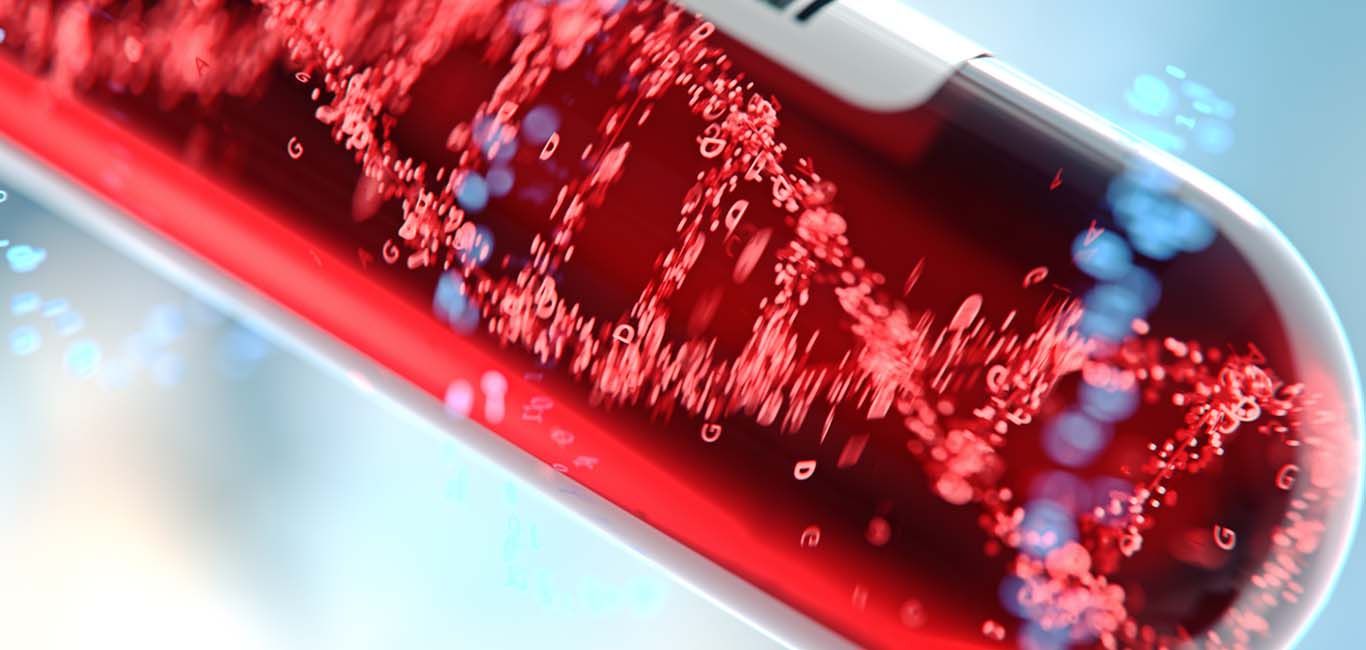
Scientists from the German Cancer Research Center (DKFZ) and the Cambridge Stem Cell Institute have jointly developed an artificial intelligence (AI) tool to analyse peripheral blood smear, a routinely used diagnostic technique. The algorithm can help health experts diagnose blood disorders quickly.
The tool can recognise and analyse white and red blood cells in microscopic images of blood smear, more accurately in lesser time than conventional methods. This could also help in reducing the need for invasive measures such as biopsy for certain blood disorders.
The researchers have made the software available as an open-source method for the research fraternity so that they can test the algorithm. The study was published in the journal Nature Communications early in August.
Current practices
Blood disorders are often identified by change in numbers and shapes of red and white blood cells. For the purpose of diagnosis, physicians examine peripheral blood smear on a slide under a microscope and analyse theses change in count and morphology (appearance, shape, size and structure) of blood cells. This type of diagnosis is straightforward.
In certain cases, evaluation by even highly experienced experts becomes difficult because the changes are unclear and affect only a few of the tens of thousands of cells seen on slide. As a result, experts could struggle to differentiate between health conditions.
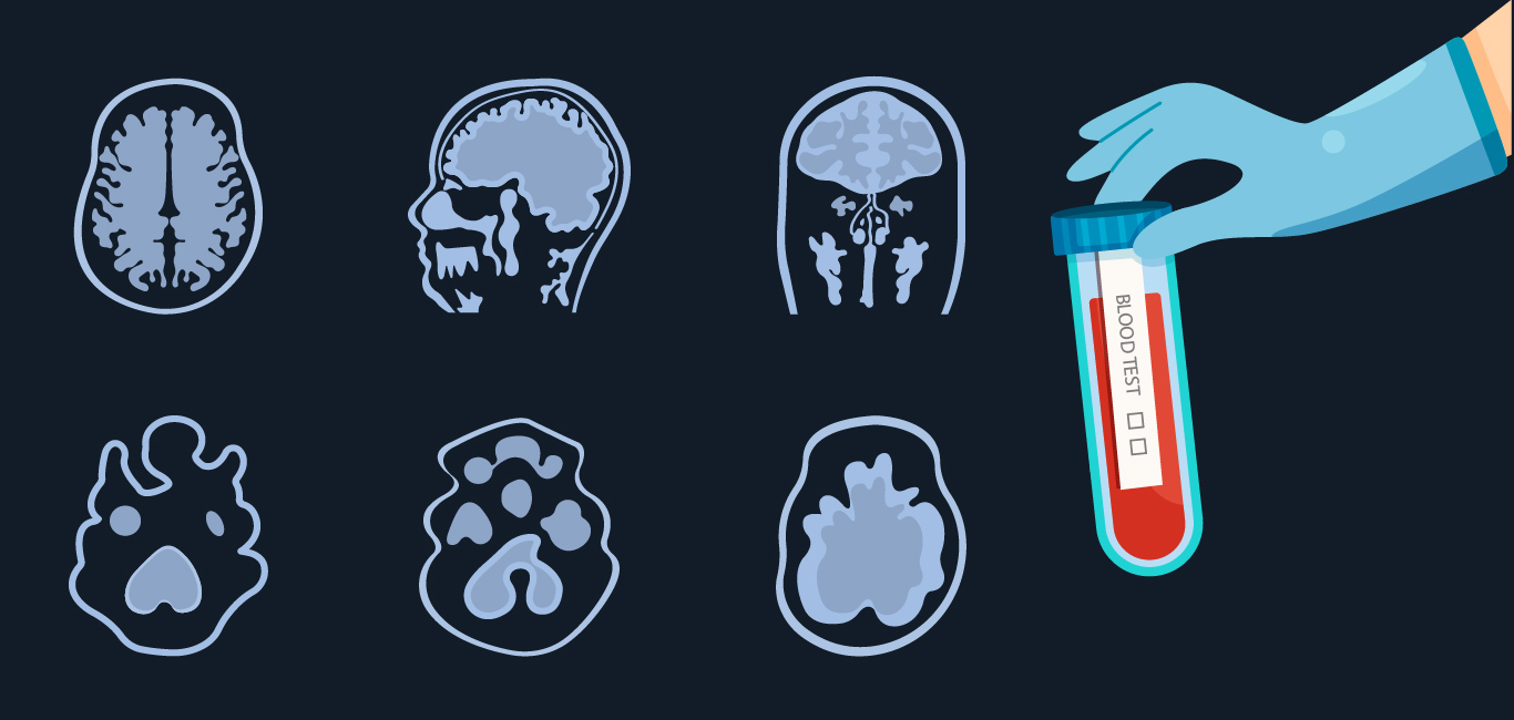
It is seen in case of people with myelodysplastic syndrome (MDS), an early form of leukemia (blood cancer). The visible changes in the peripheral blood smear in these people often resemble those of much harmless forms of anaemia (low red blood cells or haemoglobin to carry oxygen). The definitive diagnosis of MDS therefore requires more invasive procedures, such as analysis of bone marrow biopsies and molecular genetic testing.
Read More: Blood stem cell donation: Indian army braveheart duo show the way
Read More: Blood and iron: Anemia can affect your heart health
AI to the rescue
“To support specialists in these difficult diagnoses, we have developed a computer-based system ‘Haemorasis’ that automatically recognises and analyses white and red blood cells from peripheral blood smear,” explains Dr Moritz Gerstung of DKFZ.
Gerstung and his colleagues developed and trained the algorithm to recognise the structure of blood cells from the digitised images of peripheral blood smears. Thereafter, the structure and size of more than half a million white blood cells as well as many millions of red blood cells from more than 300 individuals with different blood disorders (various anaemias and genetic subtypes of MDS), was recognised by Heamorasis. These samples were obtained from Munich Leukaemia Laboratory (MLL).
“The algorithm is able to detect the shape and number of tens of thousands of blood cells in a microscopic image of the blood. This complements human capabilities, which are typically more focused on detail,” Dr Gerstung says.
External validation
Using the trained knowledge, Haemorasis is now able to diagnose blood disorders such as MDS and anaemia. It is even able to distinguish the genetic subtypes of these health conditions. In addition, the algorithm also reveals concrete correlations between certain cell morphologies and health conditions, which are often difficult to find because of the large number of cells involved.
This was demonstrated when the Haemorasis was used to analyse the peripheral blood smear samples from an independent group of 68 people from a different centre at Cambridge University Hospital. It was able to differentiate genetic subtypes of MDS from anaemia. Even the two different types of anaemia, namely megaloblastic anaemia, and iron deficiency anaemia, could be identified.
“We have now demonstrated for the first time that computer-assisted analysis of blood images is possible and can contribute to initial diagnosis,” explains Dr Gerstung.
Usage and future
Haemorasis is designed to facilitate diagnostics in hematology and can help in accurate initial diagnosis of blood disorders. This is important for identifying those people who require more invasive testing, such as bone marrow or genetic analysis.
“Automated cell analysis with Haemorasis could complement routine diagnosis of blood disorders in the future. So far, the algorithm has only been trained on specific diseases – but we still see great potential in this approach,” Gerstung says.
Read More: What blood tests reveal about infections